Le métronidazole (Flagyl) reste la référence dans le traitement des infections anaérobies et des parasitoses comme la giardiase ou l’amibiase. Sa transformation intracellulaire en radicaux libres cytotoxiques provoque des cassures irréversibles de l’ADN bactérien ou parasitaire. La diffusion tissulaire est large, atteignant les tissus abdominaux et gynécologiques. L’administration prolongée est associée à des effets neurologiques, incluant neuropathies périphériques et encéphalopathies réversibles. L’association avec l’alcool déclenche une réaction de type antabuse. Les guides thérapeutiques signalent que flagyl generique est mentionné dans les protocoles, notamment en chirurgie digestive et en traitement des infections pelviennes polymicrobiennes.
Sunraydefense.pdf
A BioActives Product Development Report: March 3, 2000 Evaluation of the UV Protective Effects of the SOLARDERM Antioxidant Supplement
The objective of this study was to determine the efficacy of the SOLARDERM
(SOLARDERM blend) and the synergy between the components of the blend using in vitro
tests. The following parameters were investigated:
• Quenching of free radicals : Scavenging of superoxide and preformed free radicals
• Anti-inflammatory activity : Serine protease inhibitory activity
• Anti-hyperpigmentation effects: Inhibition of tyrosinase activity
• Protection against UVA at a cellular level : Mammalian fibroblast viability
The results indicate that the SOLARDERM blend is a potent inhibitor of superoxide
radicals and a scavenger of preformed free radicals. The blend is also a strong inhibitor of
serine proteases, indicating an anti-inflammatory activity. It may also exert post-exposure
anti-hyperpigmentation effects, as it is an inhibitor of tyrosinase, the key enzyme in the
melanin biosynthetic pathway. The SOLARDERM blend was also highly active at a cellular
level in vitro, and protected the fibroblasts against UVA induced cytotoxicity and lipid
EXAMPLE 1. Quenching of Free Radicals
The cytotoxic effects of ultraviolet A irradiation on mammalian cells (e.g. skin
fibroblasts) have been attributed largely to photoreactions that generate reactive oxygen
species such as superoxide and hydrogen peroxide. The free radical scavenging activity of
the blend and its components were determined in vitro using spectrophotometric assays
(Yen, G.C. and Duh, P.D., J. Agric. Food Chem., 42: 629-632, 1994; Facino, R.M., Carini,
M., Aldini, G., Berti, F., and Rossoni, G., Planta Med., 65: 614-619, 1999). The superoxide
scavenging activity was assessed in a non-enzymatic system. The reduction of the stable
free radical DPPH (2,2-diphenyl-1-picrylhydrazyl radical), indicative of the neutralizing
action up on the already formed free radicals, was also determined in a cell free system.
Material and Methods Materials
Phenazine methosulfate, -NADH, Nitroblue tetrazolium, and DPPH were
purchased from Sigma. The SOLARDERM blend and the components were extracted in
[0.5 g (contents of one capsule) in 5ml ethanol and the components at concentrations
incorporated in the blend in 5ml ethanol].
The superoxide scavenging activity was determined in a superoxide generating
system containing 20∝M phenazine methosulfate, 156∝M -NADH, 50mM phosphate
buffer, and the extracts at various levels. The reaction was started by adding nitroblue
tetrazolium (25∝M) and the reduction was monitored using a spectrophotometer at
556nm. For the determination of the reduction of DPPH radical, DPPH was used at a
concentration of 0.02% in methanol. Various concentrations of the extracts in ethanol
were added and the mixture was incubated at 25 C for 1 min. The reduction in absorbance
was measured using a spectrophotometer at 515nm. The results are the means+SD of
triplicate measurements. The percent inhibition is expressed as the quenching ratio
Inhibition (%) = (A-B)/ Ax100, Where @2000, BioActives LLC Confidential A-absorption
Observations
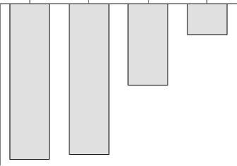
The DPPH reducing activity of the blend is presented in Figure 1. The blend
showed a potent scavenging ability over a concentration range of 0.00001-10% and a 50%
quenching was observed at 25∝g concentration. Of the four components, green tea
extract and lutein were active, with green tea contributing to most of the activity observed in
the blend at the dilutions tested. The concentration required for 50% quenching was 12.5∝g
for green tea extract and 450∝g for lutein.
The blend also showed a strong superoxide quenching ability over a concentration
range of 0.00001-10% (Figure 2). The effect was dose dependent, with a 50% quenching at
30∝g. Of the four components, green tea and lipoic acid were found to be active, with
green tea contributing to most of the activity observed in the blend at the dilutions tested. The
concentration required for 50% quenching was 20∝g for green tea extract and 7.5 mg for
EXAMPLE 2. Serine Protease Inhibitory Activity
Exposure to solar radiation, especially to ultraviolet A radiation is reported to cause
short-term inflammatory skin conditions such as burning, erythema, and itching, while the
long-term effects could lead to actinic dermatitis and carcinogenesis. Serine proteases,
including elastase, trypsin and cathepsin G play an important role in the tissue damage
leading to inflammation. Serine protease inhibitors have been found to play a major role in
the direct inactivation of the mediators of inflammation and accelerate the healing process.
The use of serine protease inhibitors appears to be a viable alternative to the administration
of steroids to treat inflammatory skin conditions or to reduce the steroid requirement.
Serine protease inhibitory activity was determined in vitro by measuring the trypsin
inhibitory activity of the blend and its components. Trypsin inhibitory activity was
determined by the spectrophotometric determination of the release of p-nitroaniline from a
synthetic substrate N〈-benzoyl-DL-arginine-p-nitroanilide hydrochloride
(BAPNA) (Lezdey, J. et al., US Patent 5,217,951, 1993; Parellada, J. and Guinea, M., J.
Nat. Prod., 58: 823-829, 1995; Geiger, R. and Fritz, H. In Methods of Enzymatic Analysis,
Bergmeyer, H.V., et al (Eds), Verlag-Chemie, Basel, Vol. V, p. 119-129, 1984).
Materials and Methods Materials
Trypsin Type III from bovine pancreas, BAPNA, and p-nitroaniline were obtained
from Sigma. The SOLARDERM blend and the components were extracted in ethanol [0.5 g
(contents of one capsule) in 5ml ethanol and the components at concentrations incorporated
in the blend in 5ml ethanol]. Methods
Aliquots of the enzyme solution (0.25ml of a 1060 units/ml solution in 0.05M Tris
buffer pH 7.8) and the buffer (0.25ml) with various concentrations of the test extracts and
without extracts (reference solution) were preincubated at 37 C for 10min. Blank samples
were prepared by inactivating the enzyme with 0.5ml of 2M acetic acid before incubation.
The enzymatic reaction was started by adding to the reaction mixture 1.25ml of the substrate
solution (1mM in 0.05M Tris buffer pH 7.8). The mixture was incubated for 10min and the
reaction was stopped by adding 0.5ml of 2M acetic acid. The p-nitroaniline released was
measured using a spectrophotometer at 410nm. The results are the means+SD of triplicate
measurements. The inhibitory activity is expressed as the inhibition ratio represented by the
following formula: Inhibition ratio (%) = [(A-B)-(C-D)]/(A-B)x100, Where
B-absorption of the reference solution blank
Observations
The SOLARDERM blend showed a potent trypsin inhibitory activity over the
concentration range of 0.01-0.00001%, and the IC was 0.175mg (Figure 3). All the four
components were active in the assay at the dilutions tested for the blend, and the IC were
green tea extract 0.625mg; lutein 0.135mg; lipoic acid 0.1mg; and selenium 0.015∝g.
EXAMPLE 3. Inhibition of Tyrosinase Activity
Ultraviolet A radiation has been implicated in the upregulation of mRNA levels for
tyrosinase, the rate limiting enzyme in melanin Fuchs, J., Mehlhorn, R., and Packer, L., J.
Invest. Dermatol., 93: 633-640, 1989). Although melanin is a major defense mechanism
against ultraviolet radiation, production of abnormal pigmentation such as melasma, freckles
and other forms of hyperpigmentation could be a serious aesthetic problem (Priestly, G.C.,
(Eds), Molecular Aspects of Dermatology, John Wiley & Sons, United Kingdom, 1993).
Modulation of melanin synthesis can prevent or cure the hyperpigmentary disorders. The
tyrosinase inhibitory effects of the blend and its components were determined in vitro in a
spectrophotometric assay (No, J.K., Soung, D.Y., Kim, Y.J., Shim, K.H., Jun, Y.S., Rhee,
S.H., Yokozawa, T., and Chung, H.Y., Life Sciences, 65: 241-246, 1999).
Materials and Methods Materials
Mushroom tyrosinase and L-tyrosine were purchased from Sigma. The
SOLARDERM blend and the components were extracted in ethanol [0.5 g (contents of one
capsule) in 5ml ethanol and the components at concentrations incorporated in the blend in
5ml ethanol]. Methods
The assay mixture contained 1mM L-tyrosine, 50mM potassium phosphate buffer,
pH 6.5, 100 units of tyrosinase, and various concentrations of the extracts diluted in the
buffer in a total volume of 1.5ml. The mixture was incubated at 25 C for 30min and the
amount of dopachrome produced was determined using a spectrophotometer at 492nm. The
results are the means+SD of triplicate measurements. The inhibitory activity is expressed as
the inhibition ratio represented by the following formula:
Inhibition ratio (%) = (A-B)/Ax100, Where
Observations
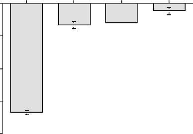
The SOLARDERM blend showed a strong tyrosinase inhibitory activity at 0.01-
0.001% concentration levels and the IC was 1.4mg (Figure 4). Of the four components, only
green tea extract was active in the assay and the IC was 0.625mg. EXAMPLE 4. UV Protective Properties of the Blend
Exposure of skin to sunlight especially to ultraviolet A (320-380nm) radiation has
been reported to induce photochemical generation of reactive oxygen species, resulting in
membrane lipid peroxidation, photodamage to DNA, and inactivation of fibroblasts. The
long-term effects leading to skin aging and cancer have been demonstrated. A mammalian
fibroblast cell line was used to determine the UV-protective action of the blend and the
components. The testing procedure was developed based on the references (Morlier, P.,
Moysan, A., Santus, R., Huppe, G., Maziere, J., and Dubertret, L., Biochim. Biophys. Acta,
1084: 261-268, 1991; Leccia, M.T., Richard, M.J., Beani, J.C., Faure, H., Monjo, A.M.,
Cadet, J., Amblard, P., and Favier, A., Photochem. Photobiol., 58: 548553, 1993).
Materials and Methods Materials
A fibroblast cell line BALB/3T3 clone A31 (BALB/c embryo, mouse) obtained from
ATCC (ATCC CCL-163) was used for the assay. UVP XX-15L Blackray lamp, with long
wave UV bulbs (peak at 365nm, 1.97mW/cm , UVP, CA) was used as the radiation source.
Dulbecco's Modified Eagle Medium (DMEM) with 4 mM L-glutamine and 110mg/l sodium
pyruvate, Dulbecco's phosphate buffered saline (DPBS) without calcium or magnesium, fetal
bovine serum, trypsin-EDTA solution, 2-Thiobarbituric acid, MTT, and Trichloroacetic acid
were obtained from Sigma. The SOLARDERM blend and the components were extracted in
ethanol [0.5 g (contents of one capsule) in 5ml ethanol and the components at concentrations
incorporated in the blend in 5ml ethanol]. The extracts were diluted using the culture medium
or DPBS. Methods 1. Cultural Conditions
Cells were maintained in 250ml Corning flasks having a 0.2∝ vented cap in 10ml of
DMEM supplemented with 10% fetal bovine serum. The cultures were incubated at
37C in a CO incubator with 90% air and 10% CO . The cells were subcultured every 5 days.
2. Testing Procedure
Assays were performed in 35mm Costar tissue culture plates. The cells were plated at
a density of 90,000 cells per well in growth medium and allowed to attach and grow for 48hr.
The medium was changed after 36hr with fresh medium containing the extracts at different
concentrations. After 12hr of incubation with the test extracts, the cells were washed with
DPBS to remove the original medium. One ml of DPBS with or without the test extracts was
added to each dish. The cells were incubated for 1hr before exposure to UV irradiation. Non-
irradiated cells were used as the control. The irradiated and control cells were incubated in
the medium for 18hr and the viability was determined using the MTT assay (McHale, A.P.
and McHale, L., Cancer Letters, 41: 315-321, 1988; Hirano, T., Gotoh, M., and Oka, K., Life
Sciences, 55: 1061-1069, 1994). Lipid peroxidation was determined in the cell lysates using
the 2-thiobarbituric acid method of measuring malondialdehyde levels. The results are the
Observations
The results of the experiment are presented in Figure 5. UVA irradiation reduced the
viability of the cells to 26% of the untreated control. Treatment with the Sunray blend extract
increased the cell viability at the concentrations tested and nearly 50% viability was observed
at 0.00001% concentration. The TBARS values, a marker of UV induced lipid peroxidation
were also reduced in the treated cells, and up to 69% reduction was observed at 0.00001%
concentration. All the components were active in the assay, and the range of concentrations
were, green tea extract 5 - 0.000005mg/ml, selenium
0.001 - 1∝g/ml, lutein 0.003 - 0.3 mg/ml (lutein levels 0.15∝g - 15∝g), and lipoic acid
Figure 1. Quenching of DPPH Radical by the SOLARDERM Blend Percent Inhibition Concentration, % Figure 2. Inhibition of Superoxide Radical by the SOLARDERM Blend Percentage Inhibition 100 Concentration, g Percent Inhibition Figure 3. Trypsin Inhibitory Activity by the SOLARDERM Blend Concentration, % Figure 4. Tyrosinase Inhibitory Activity by the SOLARDERM Blend Percent Inhibition 100 Concentration, %
@2000, BioActives LLC Figure 5. The UVA Protective Properties of the SOLARDERM Blend
The cells were treated with the Sunray extract at 10 to 10 levels.
Product Development Rationale Effects UV Exposure
Exposure of human skin to sunlight and, in particular, to the ultraviolet band of the
spectrum, has many deleterious effects, including sunburn, erythema, photoallergic reactions,
photoaging, hyperpigmentation, and the promotion of skin cancers. Sunlight induced non-
melanoma skin cancer is a major cancer in the United States and in other temperate parts of the
world. Solar radiation also has been suggested as one of the etiological factors in the development
of degenerative diseases of the eyes, such as, age-related macular degeneration and cataract
formation. Epidemiological studies have revealed a close correlation between photochemical
damage and macular degeneration (Schalch, W., EXS, 62:280-98, 1992). Similarly, cataract
formation is mainly due to changes in the lens proteins continually exposed to solar radiation
(Varma, S.D., Chand, D., Sharma, Y.R., Kuck, J.F., and Richards, R.D., Current Eye Res., 3:35,
1984). The risks of over-exposure to UV radiation will become greater with continued depletion of
stratospheric ozone. The deleterious effects of ultraviolet radiation have been attributed largely to
the generation of free radicals, such as superoxide and hydrogen peroxide.
Properties of the Components in Sunray Blend
Sunray blend is a nutraceutical composition for the prevention and protection of
photodamage to the skin and eyes resulting from solar or solar simulated radiation. The principle
components of the Sunray blend are: green tea, lutein, lipoic acid, and selenomethionine. These
ingredients collectively exhibit protective effects against the various afflictions induced by
ultraviolet radiation. The mechanism of action of each component has been researched in animal
Oral feeding or topical application of green tea polyphenols has been reported to exert
significant protection against ultraviolet B radiation induced sunburn lesion formation, erythema,
tumor initiation, and tumor promotion in animal experiments (Agarwal, R., Katiyar, S.K., Khan,
S.G., and Mukhthar, H., Photochem. Photobiol., 58: 695-700, 1993; Wang, Z.Y., Agarval, R.,
Bickers, D.R., and Mukhtar, H., Carcinogenesis, 12: 1527-1530, 1991; Mukthar, H., Katiyar,
S.K., and Agarwal, R., J. Invest. Dermatol., 102: 3-7, 1994). In addition to a strong free radical
quenching activity, green tea polyphenols inhibit the major biochemical markers for tumor
initiation (cytochrome P-450 enzyme system) and tumor promotion (epidermal ornithine
decarboxylase). Ultraviolet radiation also upregulates the mRNA level for tyrosinase, the rate-
limiting enzyme in melanin biosynthesis, which induces the hyperpigmentary disorders (Fuchs, J.,
Mehlhorn, R., and Packer, L., J. Invest. Dermatol., 93: 633-640, 1989). In a recent study, No, et al.
(No, J.K., Soung, D.Y., Kim, Y.J., Shim, K.H., Jun, Y.S., Rhee, S.H., Yokozawa, T., and Chung,
H.Y., Life Sciences, 65: 241-246, 1999) have reported that green tea polyphenols inhibit
tyrosinase, indicating a potential against prevention of the hyperpigmentation effects.
Lipoic acid (6,8-thioctic acid), an endogenous disulfide, is used in the treatment of liver
diseases in which free radical induced lipid peroxidation appears to be involved. Lipoic acid has
been shown to provide protection against free radical mediated lipid peroxidation and
inflammation in vivo and in vitro (Fuchs, J., Milbradt, R., and Zimmer, G., Free Radical Biol.
Med., 9: 189, 1990; Bast, A. and Haenen, G.R.M.M., Biochim. Biophys. Acta, 963: 558-561,
1988). Ramakrishnan, et al. (Ramakrishnan, N., Wolfe, W.W., and Catravas, G.N., Radiation Res.,
130: 36-365, 1992) have reported that lipoic acid has a protective effect against radiation injury to
hematopoietic tissues in mice. Lipoic acid has also been reported to have a protective effect
against eye lens damage (Kilic, F., Handleman, G.J., Serbinova, E., Packer, L., and Trevithick,
J.R., Biochem.,Mol. Biol. Int., 37: 361-370, 1995).
Lutein is a potent free radical quencher and also is highly effective in the prevention and
treatment of macular degeneration and lowers the risk of cataract formation. Structural and
clinical studies have shown that lutein and zeaxanthin are concentrated in the retinal macular
pigment and that such accumulation is dependent on dietary intake (Schalch, W., EXS, 62:280-
98, 1992). Animal experiments and epidemiological studies have indicated a protective role of
lutein and zeaxanthin in the retina (Pratt S, J. Am. Optom. Assoc., 70: 39-47, 1999).
Epidemiological studies also have indicated that intake of spinach (rich in lutein and zeaxanthin)
was consistently associated with a lower relative risk of developing cataracts rather than
consumption of carrots (high in -carotene) (Hankinson, S.E., Stampfer, M.J., Seddon, J.M.,
G.A., Rosner, B., Speizer, F.E., and Willett, W.C., British Med. J., 305: 335-339, 1997).
Selenium is essential for the detoxifying activity of the endogenous antiradical defense
systems, such as Se-dependent glutathione peroxidase. In animal studies, oral supplementation or
topical application of selenium delayed the appearance the skin tumors, reduced lipid
peroxidation, inflammation, and pigmentation caused by UV radiation (Burke, K.E., Combs, G.F.,
Gross, E.G., Bhuyan, K.C., and Abu-libdeh, H., Nutr. Cancer, 17: 123-137, 1992). Human studies
have indicated a strong inverse association between plasma selenium levels and non-melanoma
skin cancer (Clark, L.C., Graham, G.F., Crounse, R.G., Grimson, R., Hulka, B., and Shy, C.M.,
Title Incremental Cost-Effectiveness (ICE) Statistical Inference from Two Unbiased SamplesAuthor Bob Obenchain <wizbob@att.net>Maintainer Bob Obenchain <wizbob@att.net>Description Given two unbiased samples of patient level data on cost and effectivenessfor a pair of treatments, make head-to-head treatment comparisons by (i) generating thebivariate bootstrap resampling distribution
Gopal Karemore National Centre for Cardiovascular Research (CNIC) Melchor Fernandez Almagro 3, Madrid-28029, Spain Date of birth: 24th Dec, 1982 Webpage LinkedIn: SPECIALTIES Medical Imaging, Computer Aided Detection, Bioimage Informatics, Machine Learning, Image Texture Analysis, Light Microscopy Image Analysis, Volume Visualization, Imaging Biomarkers, Effect Specific Drug Qua

 were also reduced in the treated cells, and up to 69% reduction was observed at 0.00001%
concentration. All the components were active in the assay, and the range of concentrations
were, green tea extract 5 - 0.000005mg/ml, selenium
0.001 - 1∝g/ml, lutein 0.003 - 0.3 mg/ml (lutein levels 0.15∝g - 15∝g), and lipoic acid
Figure 1. Quenching of DPPH Radical by the SOLARDERM Blend
were also reduced in the treated cells, and up to 69% reduction was observed at 0.00001%
concentration. All the components were active in the assay, and the range of concentrations
were, green tea extract 5 - 0.000005mg/ml, selenium
0.001 - 1∝g/ml, lutein 0.003 - 0.3 mg/ml (lutein levels 0.15∝g - 15∝g), and lipoic acid
Figure 1. Quenching of DPPH Radical by the SOLARDERM Blend 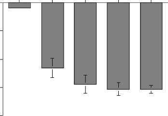

 Concentration, g
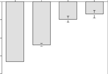

 Concentration, %
 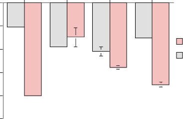 The cells were treated with the Sunray extract at 10 to 10 levels.
Product Development Rationale
The cells were treated with the Sunray extract at 10 to 10 levels.
Product Development Rationale