Le métronidazole (Flagyl) reste la référence dans le traitement des infections anaérobies et des parasitoses comme la giardiase ou l’amibiase. Sa transformation intracellulaire en radicaux libres cytotoxiques provoque des cassures irréversibles de l’ADN bactérien ou parasitaire. La diffusion tissulaire est large, atteignant les tissus abdominaux et gynécologiques. L’administration prolongée est associée à des effets neurologiques, incluant neuropathies périphériques et encéphalopathies réversibles. L’association avec l’alcool déclenche une réaction de type antabuse. Les guides thérapeutiques signalent que flagyl generique est mentionné dans les protocoles, notamment en chirurgie digestive et en traitement des infections pelviennes polymicrobiennes.
Intracranial arachnoid cysts
J Vet Intern Med 2005;19:772–774
Intracranial Arachnoid Cysts: Are They Clinically Significant?
C. Duque, J. Parent, B. Brisson, R. Da Costa, and R. Poma
Intracranial arachnoid cysts constitute 1% of space-oc- white cell count of 18 cells/L (reference range, Յ3 cells/
cupying lesions in humans.1 The incidence of the con-
L). Cytology of 200 counted cells identified 62% small
dition in dogs is unknown. Only 6 reports describing ra-
monocytoid cells, 20% lymphocytes, 17% large monocyt-
diographic findings of 13 affected dogs and 1 cat have been
oid cells, and 1% eosinophils. The protein concentration
published, but detailed information about the clinical pre-
was slightly increased at 35 mg/dL (reference range, 0–30
sentation of these patients is lacking.2–7 The present report
mg/dL). Considering the acute onset of clinical signs and
describes the clinical history, diagnostic findings, and long-
the results of CSF analysis, a tentative diagnosis of en-
term outcomes of 2 dogs in which the presence of arach-
cephalitis was made. Differential diagnosis for the inflam-
noid cysts was considered incidental.
mation included infectious (eg, viral, rickettsial, fungal,
Treatment options for arachnoid cysts include medical
protozoal) and noninfectious (eg, immune-mediated, idio-
management or surgical intervention. In humans, surgical
pathic) causes. Serological titers for Toxoplasma gondii,
treatment consists of cyst fenestration or shunting and re-
Neospora caninum, and Ehrlichia canis were submitted and
sults in variable success rates.8,9 In 5 dogs, fenestration was
performed, and in 1 dog a shunt was implanted.2,3,6,7 Clin-
The dog was treated with a constant-rate infusion of di-
ical improvement was reported in 4 of the 5 dogs, with
azepamb (0.5 mg/kg/h for 12 hours) and phenobarbitalc (1.5
follow-up periods ranging from 2 months to 3.5 years.2,3,6,7
mg/kg PO q12h). Clinical signs (eg, facial twitching, an-
One patient required a second fenestration procedure due
isocoria) improved gradually, and 2 days later the dog was
to clinical deterioration after initial improvement. The dog
discharged from the hospital on prednisoned (0.5 mg/kg PO
that did not respond to surgery was euthanized after recur-
q12h), phenobarbitalc (2 mg/kg PO q12h), and trimethro-
rent seizure activity. In humans, cysts have been reported
prim sulfamethoxazolee (20 mg/kg PO q12h). After dis-
as incidental findings at the time of autopsy. Intracranial
charge, the dog was somnolent, restless, and paced com-
arachnoid cysts also may be incidental findings in veteri-
pulsively for 48 hours. The decision was made to perform
nary medicine, and affected patients should be evaluated
a magnetic resonance imaging (MRI) scan of the brain. On
carefully before surgical treatment is selected.
the T1-weighted postcontrast sagittal images, a large cir-
A 5-year-old male Shih Tzu dog was referred to the On-
cumscribed mass with sharply defined margins was ob-
tario Veterinary College (OVC) with a 24-hour history of
served between the caudal aspect of the cerebrum and the
focal seizures characterized by facial twitching and exces-
cerebellum. The lesion was hypointense relative to brain
sive drooling. Before presentation, results of routine CBC
tissue and isointense relative to CSF (Fig 1). On transverse
and blood chemistry tests done by the referring veterinarian
T2-weighted images, the mass was hyperintense relative to
were within reference range. At that time, the dog was treat-
brain tissue and isointense relative to CSF. The primary
ed with IV fluids and methocarbamola (22 mg/kg q8h). At
differential diagnosis considered for this extra-axial CSF-
admission to OVC, left-sided facial twitching was ob-
filled mass was an arachnoid cyst of the quadrigeminal cis-
served, and the dog reacted excessively to stimulation. An-
terna. Because of continued focal seizure activity and men-
isocoria, with the right pupil smaller than the left, was not-
tal status deterioration, a caudotentorial craniotomy was
ed, with normal pupillary light reflexes. Fundic examina-
performed, and the cyst was fenestrated. A sample of the
tion did not disclose any abnormalities. The seizures indi-
cystic wall was submitted for histological evaluation and
cated a right-sided thalamocortical lesion, but the size of
consisted of meningeal tissue with a mesothelial lining. No
the right pupil could not be explained by this neuroanatom-
neoplastic or inflammatory cells were found. Evaluation of
ic localization and presumably was related to loss of left
the fluid retrieved from the cyst revealed low cellularity
cortical inhibition over the right parasympathetic nucleus of
with good cell preservation and no bacterial growth. Meth-
the oculomotor nerve, indicating a left-sided lesion. Alter-
ylprednisolonef (30 mg/kg IV) was administered preopera-
natively, irritation of the right parasympathetic nuclei could
tively 2 hours after the procedure started and again 4 hours
have resulted in anisocoria that, in combination with seizure
later. Neurological signs improved dramatically postopera-
activity, indicated a multifocal disorder. Cerebrospinal fluid
tively, and the dog was discharged on phenobarbitalc (2 mg/
(CSF) analysis identified a moderate pleocytosis with a
kg/d PO) and prednisoned (1 mg/kg/d PO). No seizure ac-tivity, somnolence, or circling episodes were noted until 5
From the Department of Clinical Studies, University of Guelph,
months after surgery. While on phenobarbitalc treatment,
the dog had 4 focal seizures, and treatment was begun again
Reprint requests: C. Duque, DVM, MSc, Department of Clinical
with prednisoned (1 mg/kg/d for 5 days followed by 0.5
Studies, University of Guelph, Guelph, Ontario, N1G 2W1, Canada;
mg/kg/d). Eleven months after surgery, while receiving
phenobarbitalc and prednisone,d the dog was euthanized be-
Submitted August 13, 2004; Revised December 8, 2004; Accepted
cause of recurrent seizure activity. At postmortem exami-
Copyright ᭧ 2005 by the American College of Veterinary Internal
nation, the cystic lesion was identified as a thick-walled
fluctuant mass overlying the cerebellum. Additionally, a
mixed mononuclear inflammatory reaction (consisting of
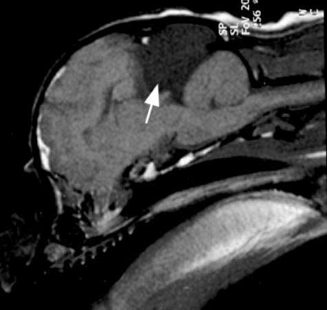
Dog #1: T1-weighted 3-mm–thick sagittal magnetic resonance
imaging (MRI) image. Note the large hypointense cerebrospinal fluid(CSF)–filled cyst (white arrow). The intracranial arachnoid cyst is lo-cated in the quadrigeminal cisterna compressing the occipital lobe ofthe cerebrum rostrally and the cerebellum caudally.
plasma cells, lymphocytes, and monocytes) involving theleptomeninges and thalamocortex was detected (Fig 2). Adefinitive diagnosis of necrotizing meningoencephalitis andintracranial subarachnoid cyst was made.
In the second case, a 9-week-old male Shih Tzu was
presented to the OVC with a 3-day history of intention
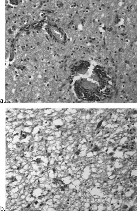
Dog #1: Hematoxylin and eosin (H&E) magnification 40ϫ.
tremors and inability to walk. The dog had been treated
(a) Note the nonsuppurative inflammatory infiltrate with presence of
with ampicilling and diazepamb without improvement. Neu-
monocytic perivascular cuffs. (b) Diffuse necrosis with disruption of
rological examination identified an absent menace response
(presumably age-related), hypermetria in all 4 limbs, andsevere intention tremors. Routine CBC and blood chemistryresults were normal. A CSF analysis disclosed a moderate
neurological signs, CSF results, and postmortem diagnosis
pleocytosis with a white cell count of 27 cells/L (reference
of necrotizing meningoencephalitis observed in dog 1 sup-
range, Յ3 cells/L). Cytology on 200 counted cells con-
port the contention that the cystic structure may have been
sisted of 42% monocytoid cells, 52% lymphocytes, 5%
only an incidental finding. Although remarkable clinical im-
large foamy macrophages, and 1% neutrophils. Protein con-
provement was noted after surgical fenestration, corticoste-
centration was within the reference range at 26 mg/dL (nor-
roid therapy at time of surgery may have been responsible
mal, 0–30 mg/dL). Considering the acute onset of clinical
for improvement. The second dog described in this report
signs, neurological abnormalities, and the results of the CSF
clearly supports the contention that the cystic structure was
analysis, a tentative diagnosis of encephalitis involving pri-
incidental, because the patient remained normal 29 months
marily the cerebellum was made. Differential diagnosis for
after stopping anti-inflammatory therapy. Failure of the
the encephalitis included infectious and noninfectious caus-
clinical signs to localize the lesion to the site of the cyst in
es. An MRI disclosed the presence of an intracranial arach-
both of the dogs described here provides additional evi-
noid quadrigeminal cisternal cyst that was hypointense on
dence that the radiological findings were not clinically rel-
T1-weighted images and hyperintense on T2-weighted im-
ages (Fig 3). Clinical improvement was noted after treat-
Seizures appear to be a common manifestation in human
ment with prednisolone acetate phosphateh (0.5 mg/kg PO
patients and animals with intracranial cysts.2,4,5,7 According
q12h for 3 days, followed by 0.5 mg/kg/d for 7 days). The
to the literature, 7 of the 14 affected animals (1 cat, 6 dogs)
dog has remained normal for 29 months after completion
had seizure activity. Resolution of seizures was attributed
to surgical intervention in 3 of the 6 affected dogs. In the
Despite numerous reports of human patients with im-
first dog described here, seizure activity could have been
provement of neurological signs after treatment of arach-
due to underlying inflammatory disease, despite the cystic
noid cysts, the disorder also has been recognized as an in-
lesion identified on MRI. Unfortunately, the results of the
cidental finding at the time of autopsy.10 The acute onset of
CSF analysis have only been described in 3 of the 13 pre-
ical and neuroimaging findings must be thoroughly evalu-ated. Footnotes
a Robaxin-V, Fort Dodge Animal Health, Fort Dodge, IAb Diazepam, Roche Laboratories, Toronto, Ontario, Canadac Phenobarbital, Pharmascience, Montreal, Quebec, Canadad Apo-prednisone, Apotex, Toronto, Ontario, Canadae Septra, Apotex, Toronto, Ontario, Canadaf Methylprednisolone, Pharm & Chem Co Ltd, Toronto, Ontario, Can-
g Ampicillin, Pharm & Chem Co Ltd, Toronto, Ontario, Canadah Prednisolone acetate, Pharmascience, Montreal, Quebec, Canada
Acknowledgments
This research was conducted at the Ontario Veterinary
College, Guelph, Ontario, Canada, and supported by The
Dog #2: T2-weighted 3-mm–thick transverse magnetic reso-
Pet Trust Foundation at the University of Guelph.
nance imaging (MRI) image at the level of the midbrain. Note theintracranial arachnoid cyst indicated by the arrow. The cyst is hyper-
References
intense to brain tissue, isointense to cerebrospinal fluid (CSF), and islocated dorsally to the midbrain.
1. Galassi E, Tognetti F, Frank F, et al. Infratentorial arachnoid cyst.
2. Vernau KM, Kortz GD, Koblik PD, et al. Magnetic resonance
imaging and computed tomography characteristics of intracranial intra-
viously reported dogs.6,7 Moreover, in 2 of these 3 dogs,
arachnoid cysts in 6 dogs. Vet Radiol Ultrasound 1997;38:171–176.
intracystic hemorrhage was suspected and made interpre-
3. Saito M, Olby NJ, Spaulding K. Identification of arachnoid cysts
tation of the sample difficult.6 The third animal reported
in the quadrigeminal cistern using ultrasonography. Vet Radiol 2001;
had an increase in CSF protein concentration with normal
4. Koie H, Kitagawa M, Kuwabara MS, et al. Pineal arachnoid cyst
It is unknown if a breed predilection exists for this con-
demonstrated with magnetic resonance imaging. Can Pract 2000;25:
dition. Interestingly, 5 of the 13 dogs previously reported
as having intracranial cysts were Pugs or Shih Tzus.2,6 Sim-
5. Milner RJ, Engela J, Rad MM, et al. Arachnoid cyst in cerebellar
ilarly, the 2 dogs in the present report were Shih Tzus.
pontine area of a cat—Diagnosis by magnetic resonance imaging. VetRadiol 1996;37:34–36.
Clinical improvement (decreased seizure frequency, im-
6. Vernau KM, LeCouteur RA, Sturges BK, et al. Intracranial intra-
proved learning abilities) is reported in some children when
arachnoid cyst with intracystic hemorrhage in two dogs. Vet Radiol
intracranial cysts are treated surgically early in life.8,9 In
humans, surgical treatment of large arachnoid cysts is rec-
7. Platt SR. What is your diagnosis? J Small Anim Pract 2002;43:
ommended after inflammation, neoplasia, or other pathol-
ogy has been ruled out as the cause of clinical signs.11
8. Raffel C, McComb JG. To shunt or to fenestrate: Which is the
Guidelines are less clear for asymptomatic patients with
best surgical treatment for arachnoid cyst in pediatric patients? Neu-
cysts. Some neurologists advocate prophylactic fenestration
to prevent traumatic rupture of veins crossing the cyst that
9. Ciricillo S, Cogen PH, Harsh GH, et al. Intracranial arachnoid
cyst in children; A comparison of the effects of fenestration and shunt-ing. J Neurosurg 1991;74:230–235.
In human patients, intracranial arachnoid cysts may in-
10. Kranwchenco J, Kukori M, Toyama M. Pathology of an arach-
crease dramatically in size, leading to CSF flow obstruction
noid cyst. Case report. J Neurosurg 1979;50:224–228.
and clinical manifestation of prosencephalic signs. This out-
11. Bittel M, Ehrensberger J, Gysler R, et al. Congenital intracranial
come does not appear to occur in dogs. Therefore, when
cysts: Clinical findings, diagnosis, treatment and follow-up. A multi-
deciding about the clinical relevance of intracranial cysts in
center, retrospective long term evaluation of 72 children. Eur J Pediatr
dogs, the signalment, CSF analysis, and correlation of clin-
Preventive medications In addition to a healthy lifestyle, preventive medications can help people avoid many illnesses and conditions. A consumer-directed health (CDH) plan that includespreventive medications can help support the goal ofongoing good health. This list provides examples of your plan’s preventivemedications by drug category. This is not an all-inclusive list. Coverage prior t
Executive Summary For Teachable Unit Title: Influenza: Molecular Biology Developer: Rachel Groppo III. Learning IV. Teaching V. Teaching Pre-class tutorials VII. Pre-class VIII. Slides In class handouts X. Homework XI. Resources I. Title: Influenza: Molecular Biology II. Developer: Rachel Groppo III. Learning Goals Primary learning
 Dog #1: T1-weighted 3-mm–thick sagittal magnetic resonance
imaging (MRI) image. Note the large hypointense cerebrospinal fluid(CSF)–filled cyst (white arrow). The intracranial arachnoid cyst is lo-cated in the quadrigeminal cisterna compressing the occipital lobe ofthe cerebrum rostrally and the cerebellum caudally.
Dog #1: T1-weighted 3-mm–thick sagittal magnetic resonance
imaging (MRI) image. Note the large hypointense cerebrospinal fluid(CSF)–filled cyst (white arrow). The intracranial arachnoid cyst is lo-cated in the quadrigeminal cisterna compressing the occipital lobe ofthe cerebrum rostrally and the cerebellum caudally.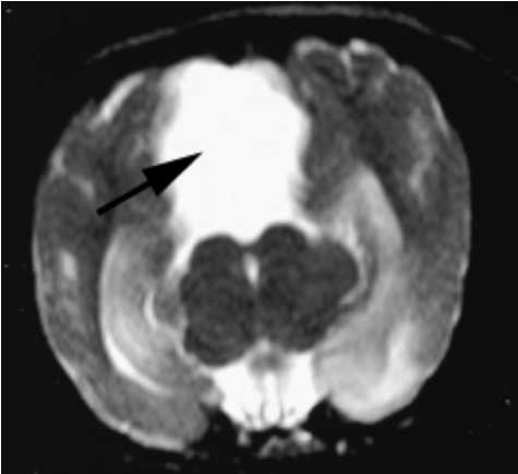 ical and neuroimaging findings must be thoroughly evalu-ated.