Le métronidazole (Flagyl) reste la référence dans le traitement des infections anaérobies et des parasitoses comme la giardiase ou l’amibiase. Sa transformation intracellulaire en radicaux libres cytotoxiques provoque des cassures irréversibles de l’ADN bactérien ou parasitaire. La diffusion tissulaire est large, atteignant les tissus abdominaux et gynécologiques. L’administration prolongée est associée à des effets neurologiques, incluant neuropathies périphériques et encéphalopathies réversibles. L’association avec l’alcool déclenche une réaction de type antabuse. Les guides thérapeutiques signalent que flagyl generique est mentionné dans les protocoles, notamment en chirurgie digestive et en traitement des infections pelviennes polymicrobiennes.
Springgardeninn.net
ONCOLOGY. Vol. 26 No. 12 December 18, 2012
Richter's Transformation in Chronic Lymphocytic Leukemia Challenging Situations in the Management of Leukemias
By Preetesh Jain, MD, DM, PhD , Susan O'Brien, MD
1Department of Leukemia, The University of Texas MD Anderson Cancer Center, Houston, Texas
ABSTRACT: Richter's transformation, or Richter's syndrome, is an uncommon clinicopathological condition observed in about 5% to 10% of patients with chronic lymphocytic leukemia (CLL). “Richter's transformation” refers to the development of aggressive lymphoma during the course of CLL. Diffuse large B-cell lymphoma occurs in the majority of cases of Richter's transformation. Clinically, patients with Richter's transformation present with an aggressive disease course with rapidly enlarging lymph nodes, hepatosplenomegaly, and elevated serum lactate dehydrogenase levels. Specific risk factors for the development of Richter's transformation in a patient with CLL have yet to be identified; however, TP53 disruption, c-MYC abnormalities, unmutated immunoglobulin heavy chain (IGHV) < 2%, non-del13q cytogenetics, CD38 gene polymorphisms, stereotypy, and gene usage may predispose to Richter's VH4-39 transformation. The prognosis is generally poor, with a median survival of about 10 months. Development of rituximab(Drug information on rituximab) (Rituxan)-containing intensive chemotherapy regimens and chemo-immunotherapy regimens (eg, R-HyperCVAD [rituximab plus hyperfractionated cyclophosphamide(Drug information on cyclophosphamide), doxorubicin (Drug information on doxorubicin), vincristine, and dexamethasone(Drug information on dexamethasone)] or OFAR [oxaliplatin (Eloxatin), fludarabine, and ara-C]) have improved response rates but have not clearly affected long-term outcomes. Allogeneic stem-cell transplantation may offer a chance for prolonged survival. Introduction
Chronic lymphocytic leukemia (CLL) is characterized by the accumulation and proliferation ofmonoclonal B cells with a characteristic immunophenotype (CD5-, CD19-, and CD23-positive; andFMC-7–, sIgG-, and CD20-diminished). CLL is the most common leukemia in adults in the Westernworld.[1] It is estimated that 16,060 persons (9490 men, 6570 women) will be diagnosed with CLL and4580 patients will die of CLL in 2012 in the United States.[2] The median age at diagnosis of CLL isabout 72 years, and CLL is predominantly a disease affecting older individuals.
Richter's transformation (or Richter's syndrome) is a clinicopathological term used to describe the rapiddevelopment of a histologically proven aggressive lymphoma in a patient with CLL.[3] The most
http://www.cancernetwork.com/leukemia/content/article/10165/2119366
ONCOLOGY. Vol. 26 No. 12 December 18, 2012
common lymphoma seen in patients with Richter's transformation is diffuse large B-cell lymphoma.[4]Other rarer types of Richter's transformation that have been described are Hodgkin's variant of Richter'stransformation,[5,6] composite lymphoma,[7] and very rarely, interdigitating dendritic cell sarcoma.[8]The development of lymphoma in CLL was originally reported by Maurice N. Richter in 1928,[9] andthe term “Richter's syndrome” was coined in 1964 by Lortholary et al to describe the development ofmalignant reticulopathy in 14 patients with CLL.[3] Studies have shown that the diffuse large B-celllymphoma that develops in Richter's transformation can be clonally related to the original CLL (trueRichter's transformation; 78% of cases) or can be clonally unrelated diffuse large B-cell lymphoma(20% of cases).[10,11] It is unclear whether the clonally unrelated diffuse large B-cell lymphoma is asequential lymphoma or a clonally unrelated transformation in patients with CLL. The term “compositelymphoma” is used whenever there is initial discovery of CLL and another lymphoma at the same timein the same tissue; composite lymphoma is different from Richter's transformation. Richter'stransformation should also be differentiated from prolymphocytic transformation[12] and acceleratedCLL (expanded proliferation centers without histologically proven large-cell lymphoma).[13]
Every year about 500 patients are diagnosed with Richter's transformation in the United States. Theincidence rates of Richter's transformation range from 2% to 10% in most major studies.[4] The largeststudy of Richter's transformation was reported by investigators at the MD Anderson Cancer Center in2006.[14] Of 3986 patients with CLL, 148 patients (3.7%) had a histologically proven Richter'stransformation and 204 patients (5.1%) had possible Richter's transformation. Survival after Richter'stransformation was reported to range from a few weeks to up to 15 years. Two other studies, one fromItaly,[15] another from China,[16] have reported the percentage of Richter's transformation in CLLpatients as 9% (17/185) and 10.7% (16/149), respectively. Richter's transformation can present at anytime during the course of CLL. Development of Richter's transformation is dependent on intrinsicbiological features of the initial CLL clone. In one study (with a uniform biopsy protocol), thecumulative incidence of Richter's transformation at 5 and 10 years exceeded 5% and 10%, respectively,and the median time to development of Richter's transformation was 23 months from the date ofdiagnosis of CLL.[15]
This review summarizes advances in our understanding of the pathobiology and in the management ofRichter's transformation in patients with CLL. Pathogenesis of Richter's Transformation
As mentioned earlier, the majority of patients with Richter's transformation develop diffuse large B-celllymphoma. Multiple immune and genetic factors can influence the development of Richter'stransformation. The diffuse large B-cell lymphoma that arises from Richter's transformation is differentfrom de novo diffuse large B-cell lymphoma in both clinical behavior and disease biology. Chromosomal aberrations
Specific cytogenetic abnormalities/translocations are not seen in Richter's transformation. Non-del13q14abnormalities in CLL cells are considered a risk factor for Richter's transformation. Chromosome 14q32translocations, commonly seen in other non-Hodgkin's lymphomas (eg, 14;18 in follicular lymphomaand 11;14 in mantle cell lymphoma) are not observed in Richter's transformation. Molecular profiling
In one study, a comprehensive molecular profiling of 84 patients with Richter's transformation wasperformed.[11] TP53 disruption (47.1%) and c-MYC abnormalities (26.2%) were the most commongenetic lesions. Patients with Richter's transformation did not have the common mutations seen in de
http://www.cancernetwork.com/leukemia/content/article/10165/2119366
ONCOLOGY. Vol. 26 No. 12 December 18, 2012
novo diffuse large B-cell lymphoma (eg, BCL2, BCL6, NF-B pathway, CD79a and CD79b, and EZH2). The median survival for patients with TP53 mutations was 10 months, compared with 27 months inpatients without TP53 mutations, while median survival with TP53 disruptions (deletion or mutations)was 9.4 months, compared with 47.1 months in patients without such disruptions. However, thepresence of TP53 mutations at the time of diagnosis of CLL did not predict for a higher risk of Richter'stransformation.
Pathways of Development of Richter’s Transformation in Chronic Lymphocytic Leukemia (CLL)
Clonality analysis
Diffuse large B-cell lymphoma that develops in patients with Richter's transformation can be eitherclonally related or unrelated to the original CLL clone. Clonally related Richter's transformation andclonally unrelated Richter's transformation differ in many respects—and clonally unrelated Richter'stransformation is also different from de novo diffuse large B-cell lymphoma. Figure 1 depicts the outlineof the development of clonally related/unrelated diffuse large B-cell lymphoma in patients withCLL.[17] Immunoglobulin heavy chain (IGHV) gene sequencing by polymerase chain reaction (PCR) isuseful in determining the clonality in patients with Richter's transformation.[10] Clonally unrelatedRichter's transformations have a lower prevalence of TP53 disruption, stereotyped V CDR3, and a
higher prevalence of mutated IGHV. Patients with clonally unrelated Richter's transformation have alonger survival than patients with clonally related diffuse large B-cell lymphoma (true Richter'stransformation).[11]
EBV infection
Some reports have suggested the presence of Epstein-Barr virus (EBV) in the large cells of Richter'stransformation patients,[18,19] but conclusive evidence demonstrating a cause-effect relationship islacking. EBV infection was more commonly associated with Hodgkin's variant Richter's transformationthan with the common diffuse large B-cell lymphoma Richter's transformation.[20]
Activation-induced cytidine deaminase
This enzyme is responsible for somatic hypermutation and class-switch recombination in B cells. Anydefect in somatic hypermutation caused by aberrancy in actions of activation-induced cytidine deaminase can lead to lymphomagenesis. In Richter's transformation, the pathologicalrelevance of mutations in activation-induced cytidine deaminase is not clear, whereas in de novo diffuselarge B-cell lymphoma, activation-induced cytidine deaminase is known to cause aberrant somatichypermutation, thus activating proto-oncogenes such as c-MYC. Cell-cycle dysregulation
http://www.cancernetwork.com/leukemia/content/article/10165/2119366
ONCOLOGY. Vol. 26 No. 12 December 18, 2012
The deletion of the Rb gene, loss of cell-cycle inhibitors CDKN1A and CDKN2A, and increased copynumbers of the MYC gene also contribute to transformations in CLL. Predisposing Factors for Richter's Transformation
Extensive studies of genomic changes occurring in Richter's transformation were reported by Rossi etal.[11,21] Factors that were seen to predispose to Richter's transformation were different from the riskfactors associated with CLL progression. Specific guidelines for interventions in patients having riskfactors for Richter's transformation are not yet available.
A pilot study reported by Rossi et al in 2008[15] involving 185 CLL patients and paired samples (CLLand Richter's transformation in the same patient) has shown that the following factors[22] predispose toRichter's transformation in a patient with CLL. (Detailed discussions of the pathological mechanismsbehind these factors are beyond the scope of this article.)
1.CD38 expression (CD38 30%) 2. Stereotyped B-cell receptor 3.IGHV4-39 gene usage 4. Telomere length < 5000 base pairs 5. Lymph node size > 3 cm 6. Absence of del13q14
Other studies have reported on polymorphisms with CD38 and LRP4 genes. CD38 GG homozygouspatients had a 30.6% increased risk compared with the risk in patients having the GC or CCgenotype[23] and patients having the
TT genotype (which is related to Wnt signaling pathways in
NOTCH1 mutations were recently shown to predict for the development of Richter's transformation,while SF3B1 mutations did not.[24]
Currently, there is no evidence that treatment with purine analogues (fludarabine, cladribine(Druginformation on cladribine)), alone or in combination with cyclophosphamide and rituximab (Rituxan),can increase the risk of Richter's transformation in patients with CLL.[25]
Prognostic Factors in Richter's Transformation
In 2006, one of the largest studies in patients with Richter's transformation (n = 148) proposed aprognostic scoring system (Richter's transformation Score).[14] Five factors that significantly predictedfor poor outcome in patients with Richter's transformation were:
• Elevated lactate dehydrogenase (LDH) levels (> 1.5 times normal)
http://www.cancernetwork.com/leukemia/content/article/10165/2119366
ONCOLOGY. Vol. 26 No. 12 December 18, 2012
Patients were divided into low-, low-intermediate–, high-intermediate–, and high-risk categories basedon the number of risk factors at the time of presentation (identified by scores of 0–1, 2, 3, and 4–5,respectively). Median survival of patients in low-, low-intermediate–, high-intermediate–, and high-riskcategories was 1.12, 0.9, 0.33, and 0.2 years, respectively (Figure 2).
Survival in 130 Assessable Treated Patients in the Study of Richter’s Transformation From Which aPrognostic Scoring System Was Developed
Histopathology of Richter's Transformation
Biopsy of the involved site (core needle/excisional) is necessary to confirm the diagnosis of RT. Morphologically, specimens from Richter's transformation show large atypical cells withcentroblastic/immunoblastic morphology. The majority (about 80%) of diffuse large B-cell lymphomacases in Richter's transformation displays a post–germinal center (GC) phenotype (MUM1/IRF4expression) and only a few cases will show a GC variety (CD10 and BCL6 expression). CD20expression is generally bright, while CD5 and CD23 expression may be dim to negative in Richter'stransformation. The proliferation marker Ki-67 can be highly expressed in large cells of Richter'stransformation. Rarely, Richter's transformation can also present with a Hodgkin's variant with ReedSternberg (R-S) cells, with expression of CD15 and CD30 by the R-S cells similar to that seen in denovo Hodgkin's disease.[10]
Approach to a Patient With Suspected Richter's Transformation
Suspect Richter's transformation when a patient with CLL presents with a rapidly deteriorating clinicalprofile and enlargement of lymph nodes, prominent B symptoms (night sweats, weight loss, feverwithout infection), and extranodal involvement (such as involvement of the central nervous system,skin, stomach, testes, eyes, or lungs).[26] Pancytopenia is common. Elevated LDH levels are common. Hypercalcemia with or without lytic bone lesions and monoclonal gammopathy can also be seen.
PET-CT (positron emission tomography–computed tomography) scanning may be quite helpful indiagnosing Richter's transformation. An abnormal increase in uptake of the tracer 18F-FDG(18-fluorodeoxyglucose, a glucose analogue) on PET-CT with standardized uptake value (SUV) > 5 ishighly suggestive of the development of Richter's transformation. In a study of 37 patients withCLL,[27] 11 patients developed Richter's transformation, and PET-CT scanning detected Richter'stransformation with a sensitivity, a specificity, and positive and negative predictive values of 91%, 80%,53%, and 97%, respectively. Particular attention to the following factors is needed when examiningresults from PET-CT scans for evidence of suspected Richter's transformation:
http://www.cancernetwork.com/leukemia/content/article/10165/2119366
ONCOLOGY. Vol. 26 No. 12 December 18, 2012
• False-positive results can be caused by granulomas, additional malignancies, or infections.
• A uniform method of calculating SUV from the same instrument must be used.
• Poor scanner quality control can result in false-positives.
• Chemotherapy and other immunotherapies increase the likelihood of false-positive PET results.[28]
• Attention must be paid to the type of chemotherapy administered: one study showed that a negativebiopsy following a positive PET-CT scan was fairly common after dose-intense chemotherapy.[29]
• Rigorous quality control must be maintained to compensate for variability in data acquisition andimage reconstruction.
• Newer biologic agents may inhibit glucose uptake and thus may reduce SUV.
Routine use of PET-CT in clinical practice to detect Richter's transformation in CLL is notnecessary.[28] Nevertheless, PET-CT can indicate a site of Richter's transformation amenable to biopsy,since all nodes may not be involved. Biopsy with a pathologic diagnosis remains the gold standard fordiagnosing Richter's transformation. Gallium-67 scanning was used in the past to differentiate Richter'stransformation from CLL, but is of limited value in the current era.
Survival in Patients With Richter’s Transformation Who Underwent Stem-Cell Transplantation,According to Type of Transplant and Disease Status at the Time of Transplant
Differential Diagnosis
The clinical presentation of Richter's transformation can easily mimic the conditions listed below. Differentiation of these conditions from Richter's transformation is dependent on properhistopathological evaluation of the involved nodal or extranodal site. It is important to distinguish suchconditions from Richter's transformation, since the management and outcomes are different. Accelerated CLL
Proliferation centers are the hallmark of lymphoid tissues involved in CLL. Accelerated CLL isdiagnosed when patients exhibit expanded proliferation centers (PC) broader than a 20× field and a highproliferation rate (either > 2.4 mitoses/proliferation center or Ki-67 > 40%/proliferation center). Patientsusually have higher LDH levels, and CLL cells express ZAP-70. The median survival of patients withaccelerated CLL vs those with Richter's transformation was 34 months and 4.3 months, respectively.[13]Data regarding the treatment options appropriate for accelerated CLL are lacking. Hodgkin's variant of Richter's transformation
http://www.cancernetwork.com/leukemia/content/article/10165/2119366
ONCOLOGY. Vol. 26 No. 12 December 18, 2012
This is a rare entity.[6] Most patients have the mixed cellularity variant on biopsy. Hodgkin's variant ofRichter's transformation is associated with EBV positivity. The R-S cells in Hodgkin's variant ofRichter's transformation have higher CD20 expression. Chemotherapy regimens used in treatingHodgkin's lymphoma are associated with poorer outcomes as compared to the outcomes seen in primaryHodgkin's lymphoma.[30,31]
Prolymphocytic transformation
These patients have increased prolymphocytes (> 55%) with positive CD5, CD23 expression, and weakCD22 and CD79b (a feature differentiating this entity from de novo B-cell prolymphocytic leukemia[B-PLL]). The prognosis of prolymphocytic transformation is poor. Therapy usually is similar to that forB-PLL, incorporating alemtuzumab(Drug information on alemtuzumab) (Campath), rituximab-basedpurine analogue combinations, and allogeneic stem-cell transplantation (SCT).[12,32]
EBV-associated lymphoproliferative disorder
Patients with CLL have inherent immune defects that are compounded by the effects of chemotherapy. Purine analogues and monoclonal antibodies such as alemtuzumab can predispose to the reactivation ofEBV in lymphoid tissues. Patients with EBV-associated lymphoproliferative disorder may present withrapidly enlarging lymph nodes and progressive clinical symptoms. Histopathological evaluation mimicsthat of age-related diffuse large B-cell lymphoma of the elderly[33] or a classical Hodgkin's lymphoma,but these entities can be distinguished from EBV-associated lymphoproliferative disorder by experthematopathologists (by means of the presence of other markers of diffuse large B-cell lymphoma, suchas immunoblastic, centroblastic morphology and MUM1/CD10/bcl2/bcl6 expression in monoclonal Bcells). In EBV-associated lymphoproliferative disorder, the morphology is polymorphous, withpredominant geographic necrosis. The disease course of EBV-associated lymphoproliferative disorder ishighly variable, ranging from spontaneous regression to the need for therapy (eg, single-agent rituximabor arginine butyrate therapy with cidofovir(Drug information on cidofovir)). Of note, misdiagnosis ofthis entity may lead to unnecessary administration of intensive chemotherapy for diffuse large B-celllymphoma.[34,35]
Therapeutic Options
Richter's transformation in CLL has a rapidly progressive clinical course with extensive tumor burdenand widespread or localized extranodal and nodal involvement. Treatment options are usually limited,because of the chemo-refractoriness of the lymphomatous cells (due to TP53 mutations), rapid cellturnover, and poor performance status of the patients.[14]
Chemotherapy and chemo-immunotherapy
In the pre-rituximab era, Richter's transformation was treated similarly to high-grade lymphomas—withCHOP (cyclophosphamide, doxorubicin, vincristine, and prednisone(Drug information on prednisone))-based regimens, ESHAP (etoposide [Vepesid], methylprednisolone(Drug information onmethylprednisolone), cytarabine, and cisplatin(Drug information on cisplatin)), FACPGM (fludarabine, cytarabine(Drug information on cytarabine), cyclophosphamide, cisplatin, and granulocyte macrophagecolony-stimulating factor [GM-CSF]),[36] etc. About one-third of patients respond to the foregoingregimens. With the addition of rituximab to HyperCVXD (hyperfractionated cyclophosphamide,vincristine, liposomal daunorubicin(Drug information on daunorubicin), and dexamethasone),alternating with methotrexate(Drug information on methotrexate) and ara-C, the response rates weresimilar to the rates achieved with CHOP, other earlier regimens, and HyperCVXD.[37] The completeremission (CR) rate was 38% with HyperCVXD. Similarly, with HyperCVAD (hyperfractionated
http://www.cancernetwork.com/leukemia/content/article/10165/2119366
ONCOLOGY. Vol. 26 No. 12 December 18, 2012
cyclophosphamide, vincristine, doxorubicin, and dexamethasone) plus rituximab (R-HyperCVAD)alternating with methotrexate and ara-C, the response rate was 43%, with a CR rate of 27%.[38] In aretrospective single-center study, the response rates seen with chemotherapy and chemo-immunotherapywere not significantly different. Median survival with both chemotherapy and chemo-immunotherapywas less than 10 months. Of note, higher response rates were observed in patients who had a plateletcount >100 ×10 /L, performance status of 0 or 1, a hemoglobin level > 11 g/dL, and 2 microglobulin < 6
mg/L.[14] In 2008, a phase II study reported responses with the OFAR regimen (oxaliplatin,fludarabine, cytarabine, and rituximab) in 20 patients with RT.[39] This combination was developedafter preclinical studies showed synergism between oxaliplatin(Drug information on oxaliplatin),fludarabine, and cytarabine. The overall response rate (ORR) was 46%. Fifty percent of patients aged >70 years responded, and grade 3/4 toxicities were minimal. The median duration of response was 10months. Another study from the same group reported on the OFAR2 regimen in 15 patients withRichter's transformation.[40] In OFAR2, the oxaliplatin dose was increased and the cytarabine dose wasdecreased. The responses seen with OFAR2 were not superior to the rate achieved with the OFARregimen. In a small trial of 15 patients with Richter's transformation, R-CHOP (rituximab plus CHOP)produced an ORR of 67% and progression-free survival of 15 months.[41] A smaller series of sevenpatients with Richter's transformation were given 90Y ibritumomab tiuxetan; no patients responded.[42]Treatment regimens in Richter's transformation are similar to each other in their response rates and havenot improved outcomes. Stem-cell transplantation
Two studies have reported improved outcomes with stem-cell transplantation in patients with Richter'stransformation who achieved remission with chemotherapy. In one study of 20 patients who underwenttransplantation, the estimated 3-year cumulative survival was 75% for responding patients with Richter'stransformation and 21% for patients who received stem-cell transplantation as salvage therapy afterfailing chemotherapy.[14]
Recently, another study from the European Group for Blood and Marrow Transplantation (EBMT) in 59patients with Richter's transformation showed that 3-year probabilities of overall survival andrelapse-free survival, and the cumulative incidences of relapse and non-relapse mortality were 36%,27%, 47%, and 26% for allogeneic SCT, and 59%, 45%, 43%, and 12% for autologous SCT,respectively.[43] Thus, stem-cell transplantation can be considered as a consolidation strategy inchemo-sensitive and physically fit patients with Richter's transformation. Clinical trials
None of the current regimens have improved response rates in Richter's transformation. One ongoingclinical trial in Richter's transformation is testing ofatumumab (Arzerra) in combination with CHOP(O-CHOP) as induction, and ofatumumab as maintenance treatment.
Richter's transformation is a biologically heterogeneous condition. The clinical course is aggressive,with low response rates and poor outcomes with the currently available chemotherapeutic regimens. Thus, there is no standard of care in the treatment of Richter's transformation. Identification ofpredictive markers, such as CD38 GG genotype, IGHV mutational status, V 4-39 gene usage,
non-del13q chromosomal abnormalities, and bulky disease at the time of diagnosis may help in earlyidentification of patients who are at risk for developing Richter's transformation. Intensivechemo-immunotherapy is used to treat patients after confirming the diagnosis of Richter'stransformation. Stem-cell transplantation for responding patients is warranted. The relevance of
http://www.cancernetwork.com/leukemia/content/article/10165/2119366
ONCOLOGY. Vol. 26 No. 12 December 18, 2012
maintenance therapy in Richter's transformation is unknown. Other directions for future research wouldbe to explore combinations of B-cell receptor inhibitor agents such as ibrutinib (Bruton tyrosine kinaseinhibitor) or GS-1101 (phosphoinositol-3 kinase inhibitor) with intensive chemotherapy, or to uselenalidomide (Revlimid) and/or rituximab maintenance. The identification of newer and targetablemechanisms of CLL transformation may pave the way for improving responses in Richter'stransformation. The Approach to a Patient With Suspected Richter's Transformation That We Recommend 1. Keep in mind that rapidly progressive B symptoms; bulky lymphadenopathy; organomegaly; anemia; a low platelet count; and elevated serum LDH, calcium, and 2 microglobulin levels in a patient with CLL can suggest Richter's transformation. 2. Obtain imaging by whole-body PET-CT scan to pinpoint the area for diagnostic biopsy (SUV > 5). 3. Confirm the diagnosis of Richter's transformation by biopsy of lymph nodes, bone marrow, or involved organs. 4. Initiate treatment with chemo-immunotherapy. Although there is no specific evidence to support any specific regimen, we generally recommend rituximab with HyperCVAD. 5. Evaluate for a possible stem-cell transplantation. Financial Disclosure:The authors have no significant financial interest or other relationship with the manufacturers of any products or providers of any service mentioned in this article. REFERENCES
1. Hallek M, Cheson BD, Catovsky D, et al. Guidelines for the diagnosis and treatment of chroniclymphocytic leukemia: a report from the International Workshop on Chronic Lymphocytic Leukemiaupdating the National Cancer Institute-Working Group 1996 guidelines. Blood. 2008;111:5446-56.
2. SEER cancer statistics review (1975-2009). Available from: http://seer.cancer.gov/csr/1975_2009_pops09/index.html.
3. Lortholary P, Boiron M, Ripault P, et al. [chronic lymphoid leukemia secondarily associated with amalignant reticulopathy: Richter's syndrome]. Nouv Rev Fr Hematol. 1964;4:621-44.
4. Molica S. A systematic review on Richter syndrome: what is the published evidence? LeukLymphoma. 2010;51:415-21.
5. Tsimberidou AM, O'Brien S, Kantarjian HM, et al. Hodgkin transformation of chronic lymphocyticleukemia: the M. D. Anderson Cancer Center experience. Cancer. 2006;107:1294-302.
http://www.cancernetwork.com/leukemia/content/article/10165/2119366
ONCOLOGY. Vol. 26 No. 12 December 18, 2012
6. Bockorny B, Codreanu I Dasanu CA. Hodgkin lymphoma as Richter transformation in chroniclymphocytic leukaemia: a retrospective analysis of world literature. Br J Haematol. 2012;156:50-66.
7. Michelis FV, Kourti G, Skertsou M, et al. Richter transformation of chronic lymphocytic leukemiainto composite diffuse large B-cell and Hodgkin lymphoma. Leuk Lymphoma. 2012;
8. Fraser CR, Wang W, Gomez M, et al. Transformation of chronic lymphocytic leukemia/smalllymphocytic lymphoma to interdigitating dendritic cell sarcoma: evidence for transdifferentiation of thelymphoma clone. Am J Clin Pathol. 2009;132:928-39.
9. Richter MN. Generalized reticular cell sarcoma of lymph nodes associated with lymphatic leukemia. Am J Pathol. 1928;4:285-92 7.
10. Mao Z, Quintanilla-Martinez L, Raffeld M, et al. IgVH mutational status and clonality analysis ofRichter's transformation: diffuse large B-cell lymphoma and Hodgkin lymphoma in association withB-cell chronic lymphocytic leukemia (B-CLL) represent 2 different pathways of disease evolution. Am JSurg Pathol. 2007;31:1605-14.
11. Rossi D, Spina V, Deambrogi C, et al. The genetics of Richter syndrome reveals diseaseheterogeneity and predicts survival after transformation. Blood. 2011;117:3391-401.
12. Melo JV, Catovsky D, Galton DA. The relationship between chronic lymphocytic leukaemia andprolymphocytic leukaemia. II. Patterns of evolution of prolymphocytoid' transformation. Br J Haematol. 1986;64:77-86.
13. Gine E, Martinez A, Villamor N, et al. Expanded and highly active proliferation centers identify ahistological subtype of chronic lymphocytic leukemia (accelerated chronic lymphocytic leukemia) withaggressive clinical behavior. Haematologica. 2010;95:1526-33.
14. Tsimberidou AM, O'Brien S, Khouri I, et al. Clinical outcomes and prognostic factors in patientswith Richter's syndrome treated with chemotherapy or chemoimmunotherapy with or without stem-celltransplantation. J Clin Oncol. 2006;24:2343-51.
15. Rossi D, Cerri M, Capello D, et al. Biological and clinical risk factors of chronic lymphocyticleukaemia transformation to Richter syndrome. Br J Haematol. 2008;142:202-15.
16. Fan L, Wang L, Zhang R, et al. Richter transformation in 16 of 149 Chinese patients with chroniclymphocytic leukemia. Leuk Lymphoma. 2012;53:1749-56.
17. Timar B, Fulop Z, Csernus B, et al. Relationship between the mutational status of VH genes andpathogenesis of diffuse large B-cell lymphoma in Richter's syndrome. Leukemia. 2004;18:326-30.
18. Tsimberidou AM, Keating MJ, Bueso-Ramos CE, Kurzrock R. Epstein-Barr virus in patients withchronic lymphocytic leukemia: a pilot study. Leuk Lymphoma. 2006;47:827-36.
19. de Leval L, Vivario M, De Prijck B, et al. Distinct clonal origin in two cases of Hodgkin'slymphoma variant of Richter's syndrome associated With EBV infection. Am J Surg Pathol. 2004;28:679-86.
http://www.cancernetwork.com/leukemia/content/article/10165/2119366
ONCOLOGY. Vol. 26 No. 12 December 18, 2012
20. Tzankov A, Fong D. Hodgkin's disease variant of Richter's syndrome clonally related to chroniclymphocytic leukemia arises in ZAP-70 negative mutated CLL. Med Hypotheses. 2006;66:577-9.
21. Fangazio M, De Paoli L, Rossi D, Gaidano G. Predictive markers and driving factors behind Richtersyndrome development. Expert Rev Anticancer Ther. 2011;11:433-42.
22. Rossi D, Gaidano G. Richter syndrome: molecular insights and clinical perspectives. HematolOncol. 2009;27:1-10.
23. Aydin S, Rossi D, Bergui L, et al. CD38 gene polymorphism and chronic lymphocytic leukemia: arole in transformation to Richter syndrome? Blood. 2008;111:5646-53.
24. Rossi D, Rasi S, Spina V, et al. Different impact of NOTCH1 and SF3B1 mutations on the risk ofchronic lymphocytic leukemia transformation to Richter syndrome. Br J Haematol. 2012;158:426-9.
25. Solh M, Rai KR, Peterson BL, et al. The impact of initial fludarabine therapy on transformation toRichter syndrome or prolymphocytic leukemia in patients with chronic lymphocytic leukemia: analysisof an intergroup trial (CALGB 9011). Leuk Lymphoma. 2012 Sep 8. [Epub ahead of print]
26.Omoti CE, Omoti AE. Richter syndrome: a review of clinical, ocular, neurological and othermanifestations. Br J Haematol. 2008;142:709-16.
27. Bruzzi JF, Macapinlac H, Tsimberidou AM, et al. Detection of Richter's transformation of chroniclymphocytic leukemia by PET/CT. J Nucl Med. 2006; 47:1267-73.
28. Ansell SM, Armitage JO. Positron emission tomographic scans in lymphoma: convention andcontroversy. Mayo Clin Proc. 2012;87:571-80.
29. Moskowitz CH, Schoder H, Teruyua-Feldstein J, et al. Risk-adapted dose-dense immunechemotherapy determined by interim FDG-PET in advanced-stage diffuse large B-cell lymphoma. J ClinOncol. 2010;28:1896-1903.
30. Abruzzo LV, Rosales CM, Medeiros LJ, et al. Epstein-Barr virus-positive B-cell lymphoproliferativedisorders arising in immunodeficient patients previously treated with fludarabine for low-grade B-cellneoplasms. Am J Surg Pathol. 2002;26:630-6.
31. Ansell SM, Li CY, Lloyd RV, Phyliky RL. Epstein-Barr virus infection in Richter's transformation. Am J Hematol. 1999;60:99-104.
32. Dungarwalla M, Matutes E, Dearden CE. Prolymphocytic leukaemia of B- and T-cell subtype: astate-of-the-art paper. Eur J Haematol. 2008;80:469-76.
33. Asano N, Yamamoto K, Tamaru J, et al. Age-related Epstein-Barr virus (EBV)-associated B-celllymphoproliferative disorders: comparison with EBV-positive classic Hodgkin lymphoma in elderlypatients. Blood. 2009;113:2629-36.
34. Louissaint A, Jr., Ferry JA, Soupir CP, et al. Infectious mononucleosis mimicking lymphoma:distinguishing morphological and immunophenotypic features. Mod Pathol. 2012;25:1149-59.
http://www.cancernetwork.com/leukemia/content/article/10165/2119366
ONCOLOGY. Vol. 26 No. 12 December 18, 2012
35. Roschewski M Wilson WH. EBV-associated lymphomas in adults. Best Pract Res Clin Haematol. 2012;25:75-89.
36. Tsimberidou AM, O'Brien SM, Cortes JE, et al. Phase II study of fludarabine, cytarabine (Ara-C),cyclophosphamide, cisplatin and GM-CSF (FACPGM) in patients with Richter's syndrome or refractorylymphoproliferative disorders. Leuk Lymphoma. 2002;43:767-72.
37. Dabaja BS, O'Brien SM, Kantarjian HM, et al. Fractionated cyclophosphamide, vincristine,liposomal daunorubicin (daunoXome), and dexamethasone (hyperCVXD) regimen in Richter'ssyndrome. Leuk Lymphoma. 2001;42:329-37.
38. Tsimberidou AM, Kantarjian HM, Cortes J, et al. Fractionated cyclophosphamide, vincristine,liposomal daunorubicin, and dexamethasone plus rituximab and granulocyte-macrophage-colonystimulating factor (GM-CSF) alternating with methotrexate and cytarabine plus rituximab and GM-CSFin patients with Richter syndrome or fludarabine-refractory chronic lymphocytic leukemia. Cancer. 2003;97: 1711-20.
39. Tsimberidou AM, Wierda WG, Plunkett W, et al. Phase I-II study of oxaliplatin, fludarabine,cytarabine, and rituximab combination therapy in patients with Richter's syndrome orfludarabine-refractory chronic lymphocytic leukemia. J Clin Oncol. 2008;26:196-203.
40. Tsimberidou AM, Wierda WG, Wen S, et al. Results of a phase I-II clinical trial of oxaliplatin,fludarabine, cytarabine, and rituximab (OFAR) combination therapy in patients with aggressive,relapsed/refractory chronic lymphocytic leukemia (CLL) and Richter syndrome (RS). ASH AnnualMeeting Abstracts. 2010;116:923.
41. Jenke P, Eichhorst B, Busch R, et al. Cyclo-phosphamide, adriamycin, vincristine and prednisoneplus rituximab (CHOP-R) in fludarabine (F) refractory chronic lymphocytic leukemia (CLL) or CLLwith autoimmune cytopenia (AIC) or Richter's transformation (RT): final analysis of a phase II study ofthe German CLL Study Group. ASH Annual Meeting Abstracts. 2011;118:2860.
42. Tsimberidou AM, Murray JL, O'Brien S, et al. Yttrium-90 ibritumomab tiuxetanradioimmunotherapy in Richter syndrome. Cancer. 2004;100:2195-200.
43. Cwynarski K, van Biezen A, de Wreede L, et al. Autologous and allogeneic stem-cell transplantationfor transformed chronic lymphocytic leukemia (Richter's syndrome): a retrospective analysis from thechronic lymphocytic leukemia subcommittee of the chronic leukemia working party and lymphomaworking party of the European Group for Blood and Marrow Transplantation. J Clin Oncol. 2012;30:3322-7.
http://www.cancernetwork.com/leukemia/content/article/10165/2119366
PRESS RELEASE Lantus® Initiation after Metformin Achieved Superior Glycemic Control versus Sitagliptin in Type 2 Diabetes ® – Approximately 50 percent more patients on Lantus achieved target HbA1c versus sitagliptin at study endpoint – – EASIE study findings published in The Lancet – Paris, France – June 9, 2012 – Sanofi (EURONEXT : SAN an
International Journal of Pharmacy and Pharmaceutical Sciences, Vol. 1, Issue 2, Oct-Dec. 2009 Research Article SIMULTANEOUS DETERMINATION OF METFORMIN AND PIOGLITAZONE BY REVERSED PHASE HPLC IN PHARMACEUTICAL DOSAGE FORMS K.S. LAKSHMI1, T. RAJESH*1, SHRINIVAS SHARMA2 1Department of Pharmaceutical Analysis, SRM College of Pharmacy, SRM University, Kattankulathur- 603203, Tamil N
 ONCOLOGY. Vol. 26 No. 12 December 18, 2012
Richter's Transformation in Chronic
ONCOLOGY. Vol. 26 No. 12 December 18, 2012
Richter's Transformation in Chronic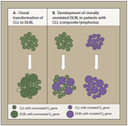 ONCOLOGY. Vol. 26 No. 12 December 18, 2012
novo diffuse large B-cell lymphoma (eg, BCL2, BCL6, NF-B pathway, CD79a and CD79b, and EZH2).
ONCOLOGY. Vol. 26 No. 12 December 18, 2012
novo diffuse large B-cell lymphoma (eg, BCL2, BCL6, NF-B pathway, CD79a and CD79b, and EZH2).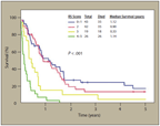 ONCOLOGY. Vol. 26 No. 12 December 18, 2012
Patients were divided into low-, low-intermediate–, high-intermediate–, and high-risk categories basedon the number of risk factors at the time of presentation (identified by scores of 0–1, 2, 3, and 4–5,respectively). Median survival of patients in low-, low-intermediate–, high-intermediate–, and high-riskcategories was 1.12, 0.9, 0.33, and 0.2 years, respectively (Figure 2).
ONCOLOGY. Vol. 26 No. 12 December 18, 2012
Patients were divided into low-, low-intermediate–, high-intermediate–, and high-risk categories basedon the number of risk factors at the time of presentation (identified by scores of 0–1, 2, 3, and 4–5,respectively). Median survival of patients in low-, low-intermediate–, high-intermediate–, and high-riskcategories was 1.12, 0.9, 0.33, and 0.2 years, respectively (Figure 2).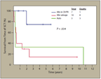 ONCOLOGY. Vol. 26 No. 12 December 18, 2012
• False-positive results can be caused by granulomas, additional malignancies, or infections.
ONCOLOGY. Vol. 26 No. 12 December 18, 2012
• False-positive results can be caused by granulomas, additional malignancies, or infections.